-40%
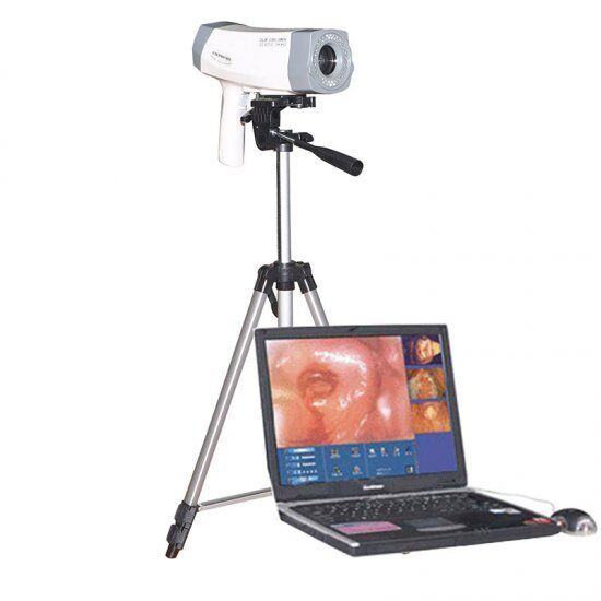
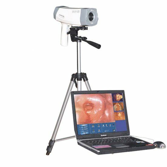
Digital Video Electronic Colposcope 480,000 Pix Camera Software&Tripod US
$ 271.91
- Description
- Size Guide
Description
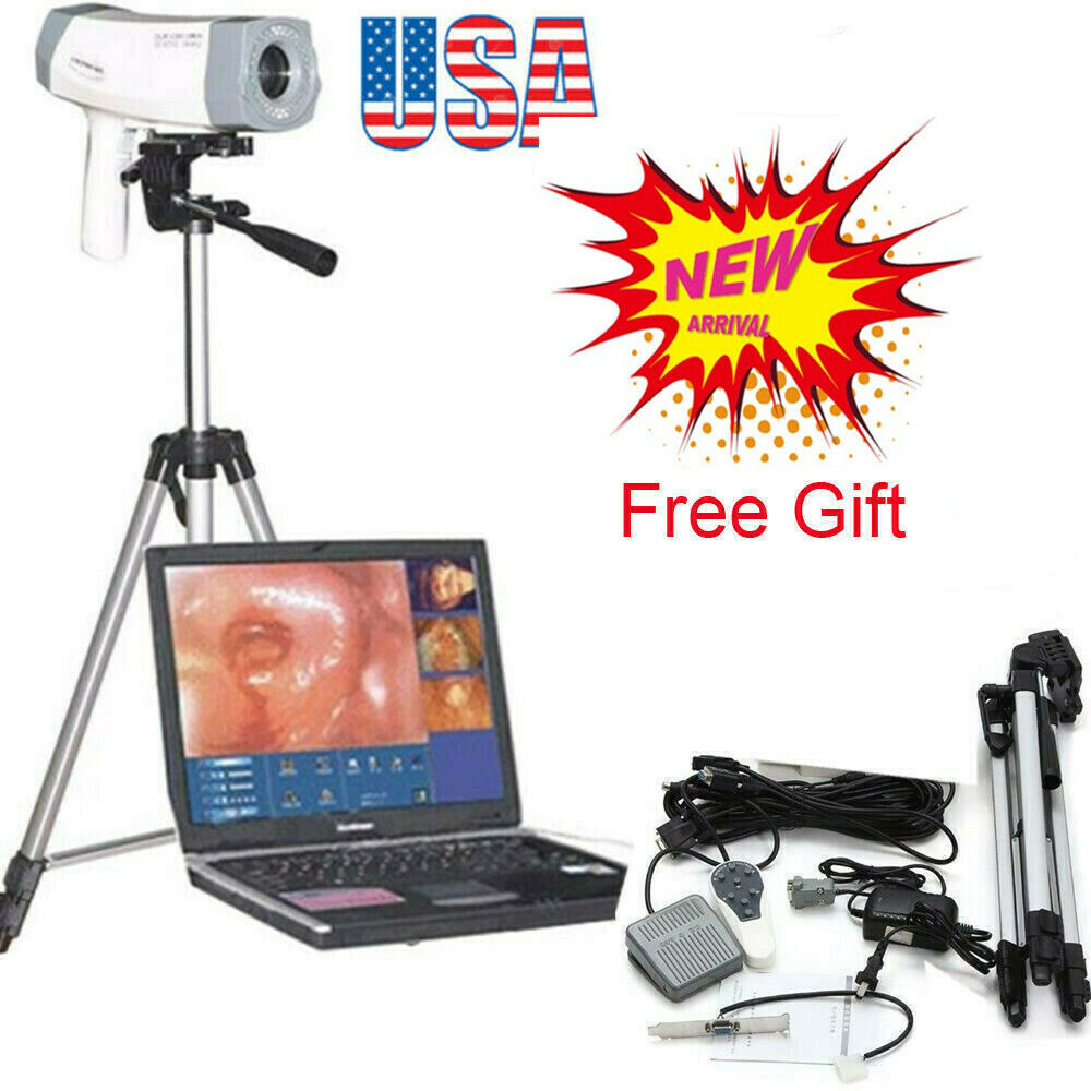
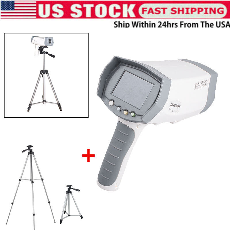
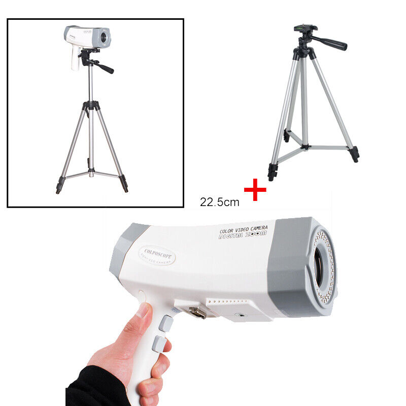
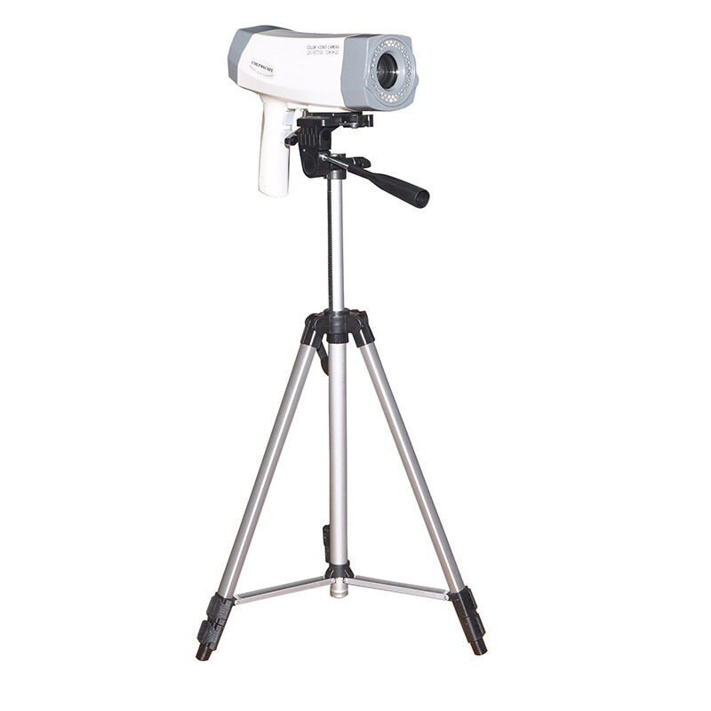
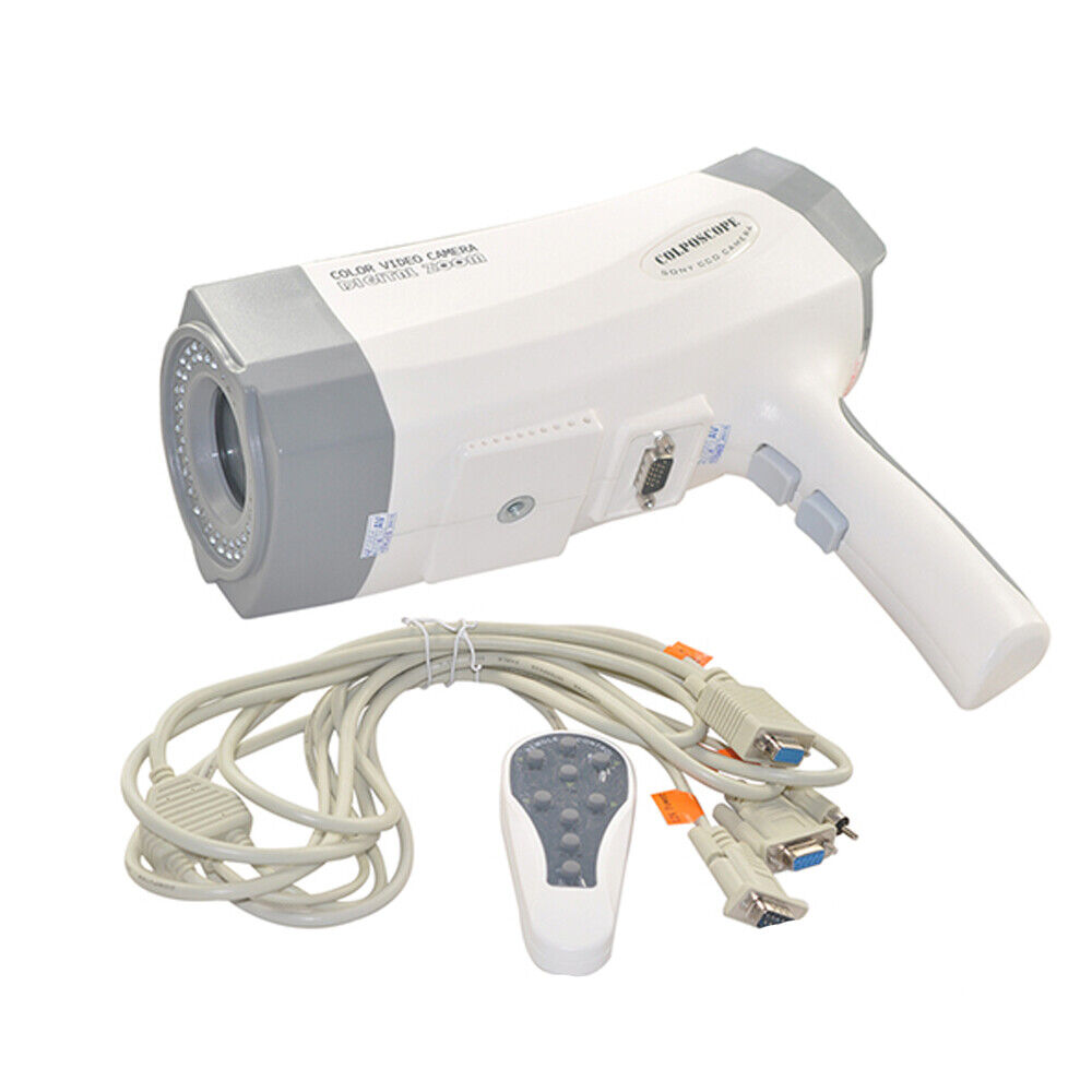
Description:RCS-400 digital electronic colposcope is applicable to detect vulva, vagina, cervical disease diagnosis and inspection, especially the cell falls off, cytology positive, macroscopic invisible lesions area which is hard to confirm.
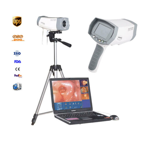
With the colposcope during Biopsy it can greatly improve the rate of the positivity. It is an extremely effective method with the colposcope while examing the early cerical cancer diagnosis cytology. It consists of video collection system, microcomputer, Colposcope software. It is mainly used for processing, saving and transmitting the medical image which is capturing by the colposcope. Since the Colposcope is an integrated image processing system, which is the newly computer aided colposcope, so it is able to show the maximum performance of the colposcope and highly meets the development of digital electronical colposcope with the DSP technology requirement in the future.
Features:
- Lines doesn't distort or overlap.
- Image quality can reach five gra
de of damage system or above.
- Have the magnetic interference function which can avoid distortion.
Application:
Vaginalitis, cervical ectropio, cervicitis, especially examining in the early diagnosis of cancer
Specifications:
-
Video
matrices
: SONY 480,000 pixels, import high-definition digital color camera
-
1/4 inch color digital CCD
-
Minimum illumination: 0.02Lux
-
Lens SNR 50DB
-
Lens Focus distance150
-350mm
, amplify or narrow in the real time
-
DSP dynamic and automatic focus system, cooperating with the manual focus adjustment
-
Magnification1-126, filter technique
-
Line-of-site:
2.5mm
-20mm
-320mm
-
Camera resolution: ≥480TVL
-
Handle: Automatic adjustment
-
Image Card: Video capture card
-
Software system: WINDOWS XP operating system, static, dynamic image continual capturing and replaying, many images in one screen; easy for measuring, calculating, copying, editing freely, printing multi-format report, including the diagnosis database, offering the case analysis of reference
Function and Characteristics:
-
High-quality 1/4 inch color digital optical zoom lens, 27 times optical zoom, 600.000 pixels providing whole screen high-definition images
-
DSP dynamic auto focus system, 8 times digital zoom cooperating with the manual focal length adjustment, super near camera, can get the clear image far from
0.1m
-
AWB way internal automatically measuring light, brightness auto adjustment and electronic shock-proof function, dynamic balance of focusing around
-
Quick focus CCD technique makes the clinical examination, observing, operating more conveniently
-
With the high speed and professional computer, it is able to realize integrated management for freezing, capturing, saving, analysis, printing, operating the images of lesions
-
High brightness light design to improve your observing effect
-
Patented multi-point ring brightness LED light source and color temperature has increased 60% compared to the traditional halogen light, life can reach to 100,000 hours, reproduce more real colors of the organization, eliminate the phenomenon such as non-uniform due to color temperature and purity and blurred image magnification
-
Sole Filter function to make sure to see the vein clearly with less loss of the light
-
Image flare subtractive radiography, shows subcutaneous vessels form more clearly, identify extremely tiny detail more easily, special effect of observing the cervix, genital blood
-
Continuous capturing static images function, and it can browse the dynamic dozens of image
-
Capacity of cine loop and record continuously for different frequencies dynamic image in sampling
-
Image database can be edited at any time, and dynamic review dozens of images
-
Tens of image enhanced functions to improve the checking rate of micro-focus
-
Capacity of internet report, and transmit the data through the internet to make an long-distance consultation and share of the information
